The clinical workflow
Follow the sample from imaging and targeted acquisition to tissue measurements and spatial interpretation.
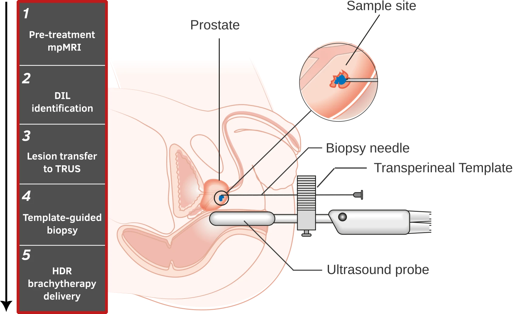
From the target to the specimen.
MRI identifies the target. Ultrasound and a transperineal template guide tissue acquisition. BiopsyContext reconstructs where the core likely sampled, retaining uncertainty when relating it to anatomy and dose.
Targeting & guidance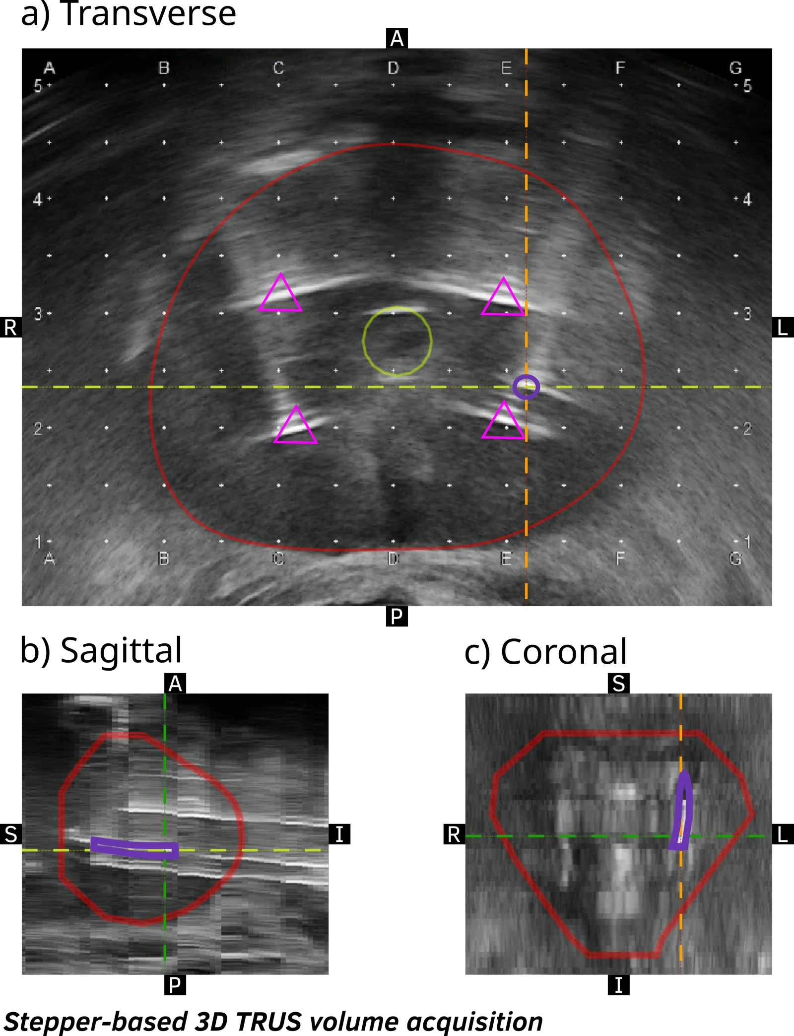
Keep the acquisition context.
The image planes connect the needle and reconstructed core to the surrounding anatomy. That geometry is the starting point for sampling and dose estimates.
Locate the sample
Connect acquired tissue with its reconstructed position in imaging space.
Preserve the uncertainty
Evaluate plausible sampling locations instead of treating a nominal path as exact.
Interpret the tissue
Relate sampling and dose descriptors to pathology or molecular measurements, with the limits of that relationship explicit.