What did the biopsy sample?
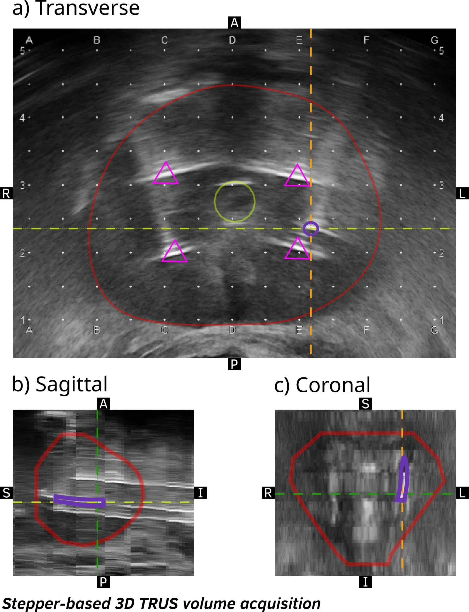
Estimate likely target sampling along a biopsy to assess targeting and give downstream tissue measurements anatomical context.
Research connections
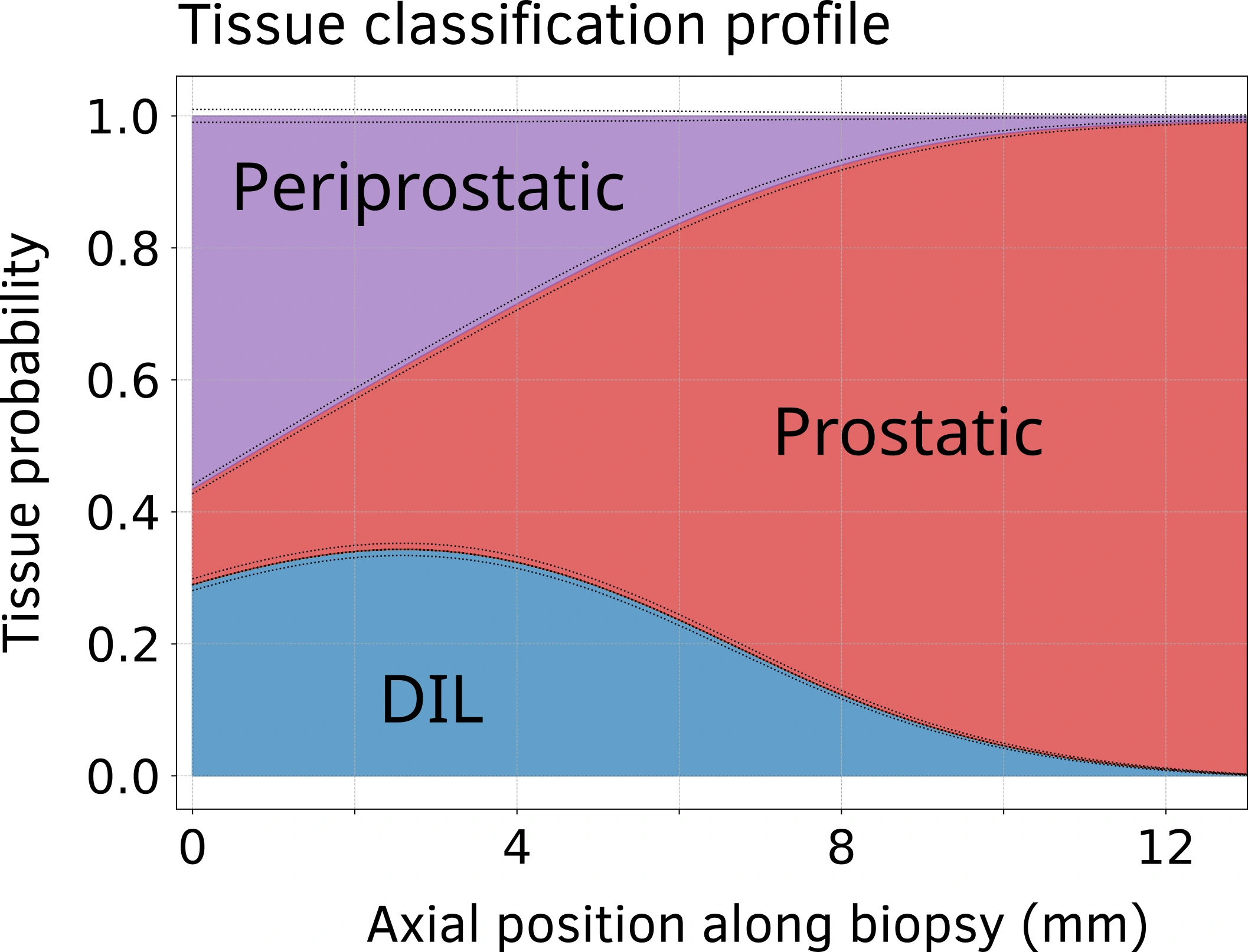
A core can cross several tissue regions.
Sampling probabilities retain those transitions along the specimen, including where it was most likely to intersect an MRI-defined lesion.
Interpretation. These probabilities describe sampling of imaging-defined structures, not the probability of cancer.
Test plausible locations, not just one path.
Shift the reconstructed core within its assumed location uncertainty, then count how often each position falls inside the lesion or surrounding tissue. Repeating this produces the sampling-probability profile.
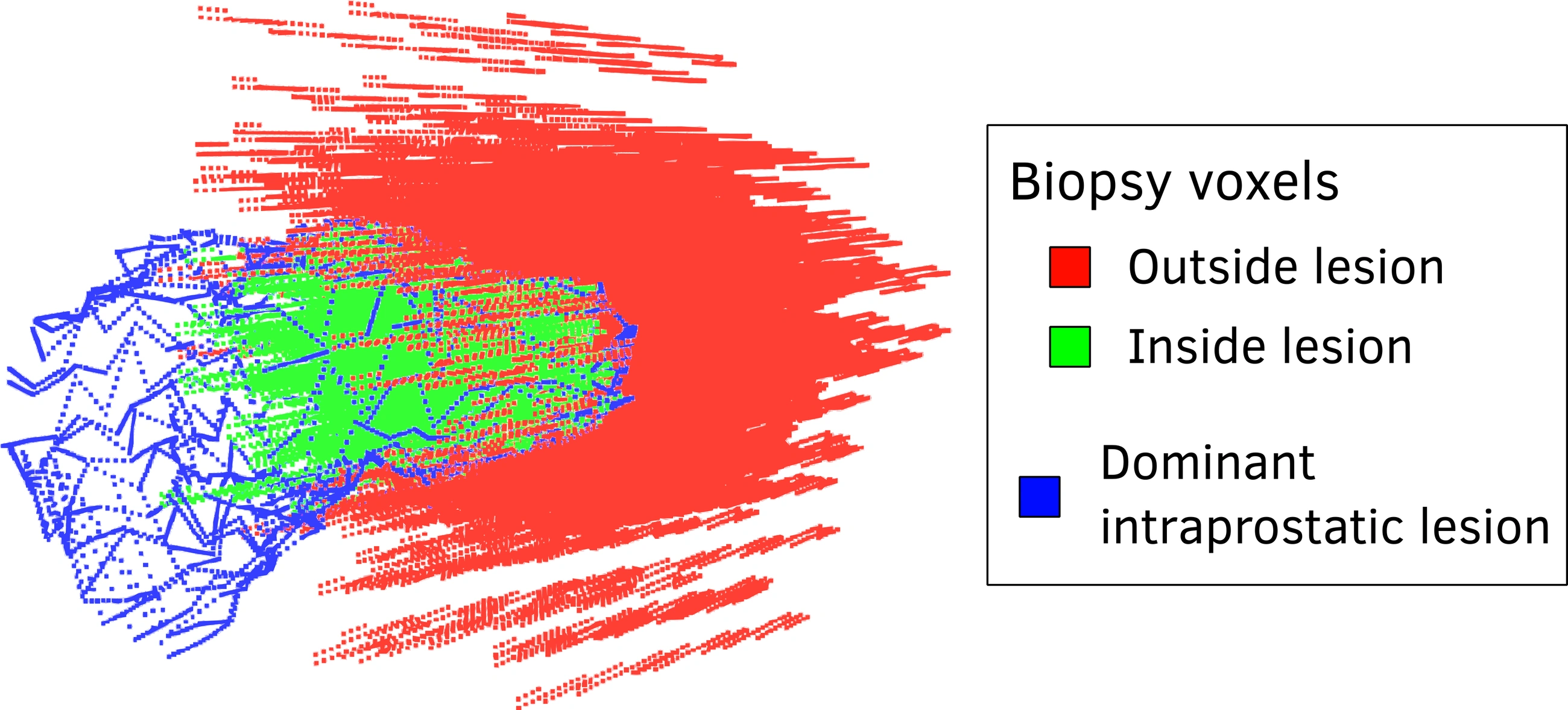
Reconstruct the sample. Test plausible positions.
The study linked 51 biopsy tracks from 15 patients to MRI anatomy containing 26 lesions. Ten thousand simulated shifts per track estimated sampling probabilities along the core.
Keep the context along the tissue.
A whole-core label can hide a target-enriched segment or a transition into surrounding tissue. Position-specific probabilities retain that context for pathology and molecular measurements.
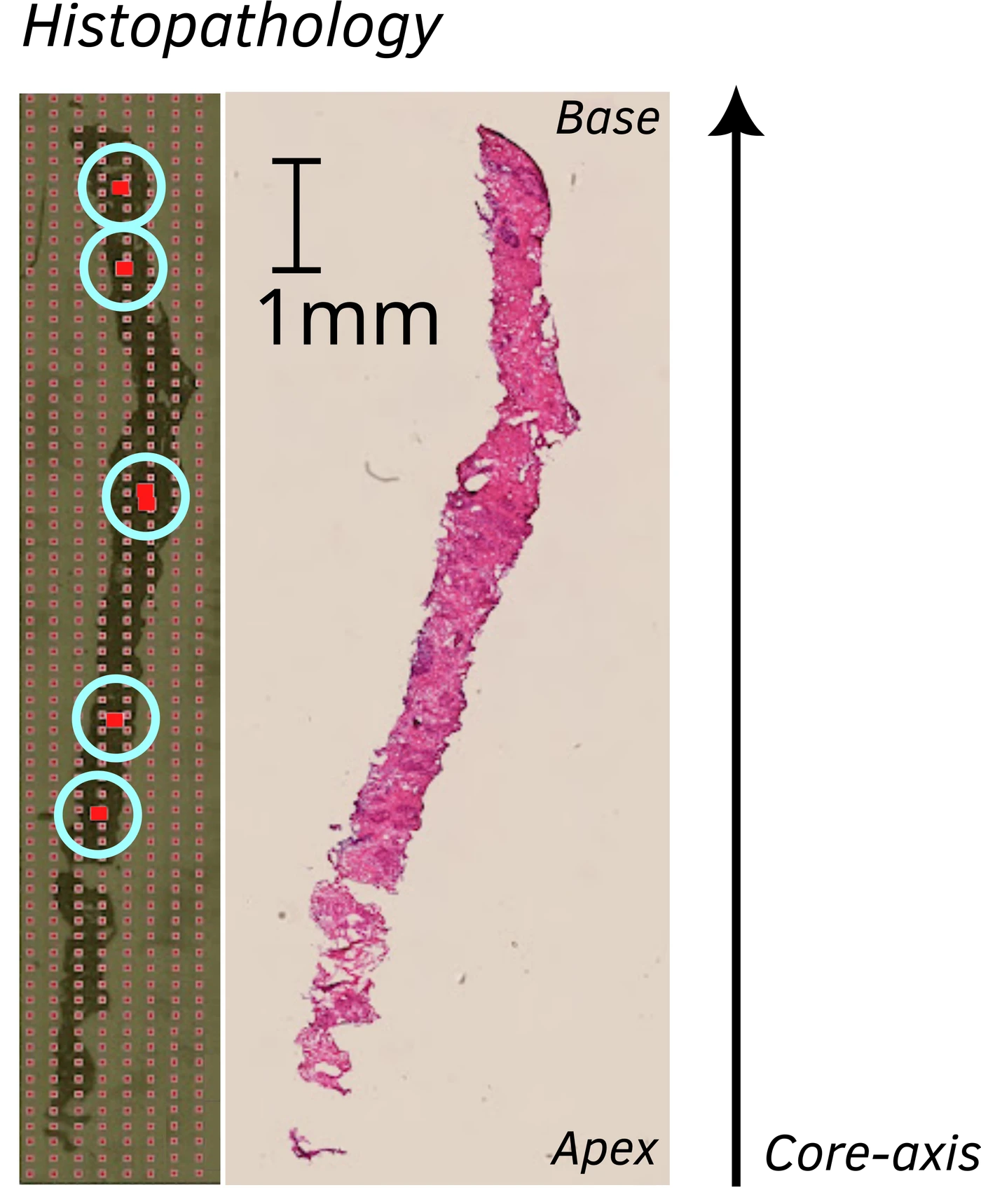
Sampling varies within each core.
The median core-average lesion-sampling probability was 0.24; the median peak was 0.46. Local peaks reveal likely target sampling that a whole-core average can obscure.
Compare sampling with pathology.
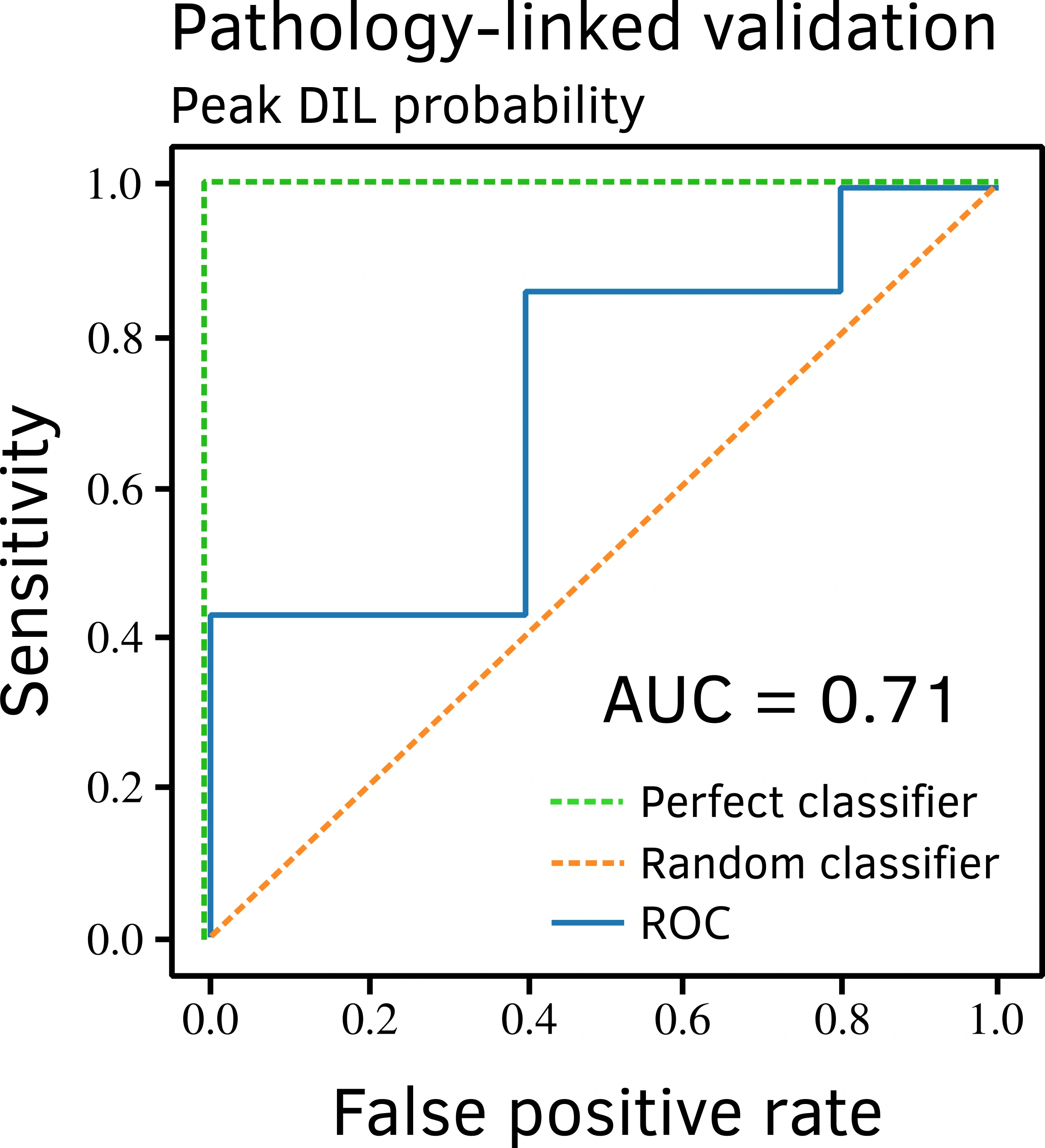
Peak lesion-sampling probability was compared with malignancy in 12 pathology-linked cores. The receiver operating characteristic (ROC) curve summarizes this exploratory comparison, with an area under the curve of 0.71.
Study scope. A small exploratory subset, not clinical validation.
From targeting QA to tissue analysis.
Use sampling profiles to assess acquired cores, compare candidate paths and give laboratory measurements anatomical context. Larger prospective studies with reliable specimen matching are needed to test these applications.